
Named after Robert William Smith after he described the fracture in A Treatise on Fractures in the Vicinity of Joints. Displaced fractures without satisfactory reduction should be seen urgently. Non-displaced fractures can be seen in orthopedic follow-up within 10 days. The position of immobilization is under debate however, the wrist should be kept in a position of function and slightly dorsiflexed and neutral. 2Ī sugar-tong or double sugar-tong splint should be applied. Reduction is achieved with longitudinal traction, over-exaggeration of the deformity (dorsiflexion), and replacement of the dorsally and proximally displaced fracture fragment into normal anatomic position. It is commonly associated with ulnar styloid process fractures, scapholunate dissociations, and injuries to the triangular fibrocartilage complex (TFCC). There is significant tension on the volar aspect of the radius and compression on the dorsal aspect. It occurs as a result of forced extension of the wrist, generally from a fall on an outstretched hand ( FOOSH). Originally described by Abraham Colles in 1814 after noticing a distinct fracture pattern in low energy injuries in elderly people, this fracture is typically dorsally displaced and angulated. However, it was first described in 1842 by Cooper, 92 years before Galeazzi reported his results.Author: Jason Brown, MD (EM Resident Physician, University of Maryland) // Edited by: Alex Koyfman, MD (EM Attending Physician, UT Southwestern Medical Center / Parkland Memorial Hospital, and Brit Long, MD EM Chief Resident at SAUSHEC, USAF)

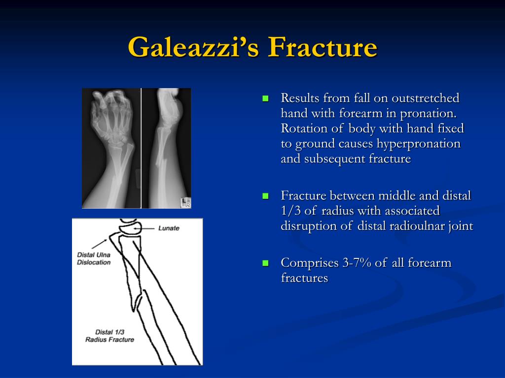
The Galeazzi fracture is named after Ricardo Galeazzi (1866–1952), an Italian surgeon at the Instituto de Rachitici in Milan, who described the fracture in 1934. They are associated with a fall on an outstretched arm. Although Galeazzi fracture patterns are reportedly uncommon, they are estimated to account for 7% of all forearm fractures in adults. Galeazzi fractures account for 3-7% of all forearm fractures. However, in skeletally immature patients such as children, the fracture is typically treated with closed reduction. Nonsurgical treatment results in persistent or recurrent dislocations of the distal ulna. It has been called the "fracture of necessity," because it necessitates open surgical treatment in the adult.

Galeazzi fractures are best treated with open reduction of the radius and the distal radio-ulnar joint. Treatment Galeazzi fracture after surgical fixation The deforming muscular and soft-tissue injuries that are associated with this fracture cannot be controlled with plaster immobilization. Īfter the injury, the fracture is subject to deforming forces including those of the brachioradialis, pronator quadratus, and thumb extensors, as well as the weight of the hand. If the fall is on the outstretched hand with forearm in pronation, the dislocation is dorsal, and if forearm is in supination at the time of injury, the dislocation is volar. The dislocation of ulnar head in Galeazzi fracture dislocation may be dorsal (commoner) or volar (rare) depending on the mechanism of injury. Galeazzi fractures are sometimes associated with wrist drop due to injury to radial nerve, extensor tendons or muscles. Injury to the AIN can cause paralysis of the flexor pollicis longus and flexor digitorum profundus muscles to the index finger, resulting in loss of the pinch mechanism between the thumb and index finger. A purely motor nerve, the AIN is a division of the median nerve. Anterior interosseous nerve (AIN) palsy may also be present, but it is easily missed because there is no sensory component to this finding.

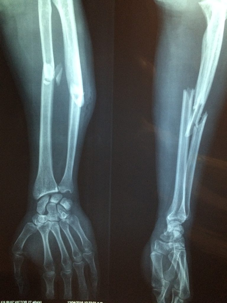
Forearm trauma may be associated with compartment syndrome. This injury is confirmed on radiographic evaluation. Pain and soft-tissue swelling are present at the distal-third radial fracture site and at the wrist joint. It classically involves an isolated fracture of the junction of the distal third and middle third of the radius with associated subluxation or dislocation of the distal radio-ulnar joint the injury disrupts the forearm axis joint. The Galeazzi fracture is a fracture of the distal third of the radius with dislocation of the distal radioulnar joint. Arrow points at the dislocated ulnar head Medical condition Galeazzi fracture-dislocation


 0 kommentar(er)
